By Carmen Willings teachingvisuallyimpaired.com Updated 10/24/2015
In order to determine how well a child is able to use their vision, an optometrist or ophthalmologist will formally assess the child's vision. Vision tests check many different functions of the eye. The tests measure a persons ability to see details at near and far distances, check for gaps or defects in the field of vision, and evaluate the ability to see different colors.
Amsler grid test
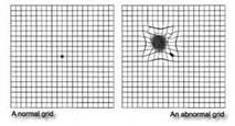
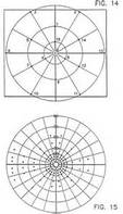
The Amsler grid test checks for macular degeneration, a disease that causes loss of vision in the center of your visual field. The test uses a 4 inch square chart with straight lines that form boxes. The grid has a black dot at the center. The chart is held about 14 inches from the face. The student must report when they cannot see the black dot; they see a blank or dark spot; or the lines in the grid look wavy, blurred or curved instead of straight.
Biomicroscope (Slit-Lamp)
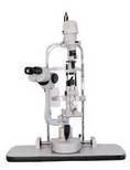
A Biomicroscope, or slit lamp, is an instrument used to view the cornea, iris, lens, and vitreous (the parts of the eye that bend light). It sends out an intense beam of light as it views the eye. It provides a magnified view of the eye structures in detail.
Confrontation Test
The confrontation test is done by having the student focus on the health professionals nose. He or she will slowly move a finger or hand from the outer edge of the visual field toward the center and from the center toward the edge through all areas of the visual field.
Keratometer
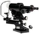
The keratometer, also known as an ophthalmometer, is a diagnostic instrument that measures the shape of the cornea and reflection of the anterior surface of the cornea. It is primarily used to diagnose the presence of astigmatism. It can also be used in surgery using the light as a point of focus for the patient.
Ophthalmoscope
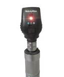
The ophthalmoscope is an instrument used to view the optic disk, retina, macula, and choroid in the back of the eye. It is typically used in routine exams and is important in determining the health of the retina and the vitreous humor. There are two types: the direct ophthalmoscope and the indirect ophthalmoscope.
Perimetry testing
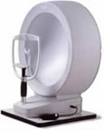
Perimetry testing uses a machine that flashes lights randomly at various points in the visual field. The student will be asked to look inside a bowl-shaped instrument called a perimeter. While the student stares at the center, lights will flash, and they will be asked to press a button each time they see a flash. A computer records the location of each flash and whether the student pressed the button when the light flashed in that location.
Refraction
Refraction is a test that measures the eyes' need for corrective lenses (refractive error). It is typically done after the visual acuity test. Refractive errors, such as nearsightedness or farsightedness, occur when light rays entering can't focus exactly on the nerve layer (retina) at the back of the eye. The eyes will be dilated with eyedrops prior to the test. Using a retinoscope, the health professional may shine light into the eyes. A series of trial lenses will be placed in front of the eyes and adjusted until the light rays are properly focused on the retina. The phoropter is another tool that can be used to measure the refractive error in the eyes.
Tonometer and Tono-pen
The tonometer is used to ensure a persons optic nerves are healthy by measuring the intraocular pressure. The small disk is placed lightly on the anesthetized eye for a brief period. The tono-pen uses a puff of air to measure eye pressure. Measurements that are higher than normal can be a sign of early glaucoma or retinal detachment.
Visually Evoked Response/Potential (VER/VEP) Test
A computerized recording of electrical activity in the vision portion of the brain that result from stimulating the retina with light flashes. These tests are used to evaluate vision functioning in the retina-to-brain nerve pathway and can be helpful in diagnosing Cortical Visual Impairment (CVI).
Tangent Screen Test
The tangent screen test uses a black screen with concentric circles and lines leading out from a center point (like a bull's-eye). Sitting 3 ft to 6 ft away from the screen, the student will be asked to cover one eye while fixing their gaze on a target point marked on the screen. Test objects of various sizes at the tip of a wand are then moved inward from the outer edge of the screen toward the center. The student will signal when he can see the object, and that point is marked on the screen. The points on the screen that objects are seen are connected to provide an outline of the visual field.
Visual Acuity Tests
Visual acuity tests are the most common tests used to assess vision. They measure the eye's ability to see details at near and distance. The tests involve reading letters, numbers or symbols of different sizes on an eye chart. Usually, each eye is tested by itself. Then, both eyes may be tested together, with and without corrective lenses if they're worn.
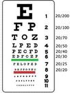
The Snellen test checks the persons ability to see at distances, measured at 20 feet. It is a wall chart that has several rows of letters. The letters on the top row are the largest and those on the bottom row are the smallest. The E chart tests the vision of children and people who cannot read, but can verbally respond to questions. It is similar to the Snellen chart in that it has several rows. Each row contains only the letter E, but in different positions. The student will be asked to point in the same direction as the lines of the E.
There are guidelines for standardized eye chart design as the design can significantly affect visual acuity scores. For school age children and older, the following guidelines should be followed: 1. Optotypes (The name for the picture, symbol, letter, or number the child is to identify) should be of approximate equal legibility. 2. Each line on an eye chart should have the same number of optotypes. 3. Horizontal spacing between optotypes should be equal to the width of the optotypes on a line. 4. Vertical spacing between lines hsould be the height of the optoypes in the next line down. 5. The size of optotypes should progress down the chart by 0.1 log units between rows. 6. Optoptypes should be black on a white background under good lighting conditions.
Visual Field Test
Visual field tests are used to check for gaps in your side (peripheral) vision. Your complete visual field is the entire area seen when your gaze is fixed in one direction. The complete visual field is seen by both eyes at the same time, and it includes the central and peripheral visual fields.
Unique Testing for Infants and Toddlers
Infants and toddlers will not be able to read typical visual acuities charts, nor will students with significant cognitive impairments. This can make the assessment more challenging and will also make it difficult to get a precise acuity. For that reason, the doctor may use other tests or measurement techniques in order to gather information about the child's vision. Some of these tests are listed below:
Confrontation field test
A screening method for detecting gross visual field loss. An interesting visual target is slowly moved inward toward the eyes of a child from various locations (above; below; from the sides) as the child looks straight ahead. The examiner watches to see at what point the child notices the visual target coming from each direction and can determine if there seem to be areas of the child's visual field in which the child cannot see.
Cover Test
Procedure for detecting eyes that are not in alignment, by watching for eye movements as a child's eyes are covered and uncovered one at a time.
Photoscreening
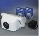
A screening procedure in which a special camera takes photographs of the eyes. The resulting pictures can identify eyes that may be nearsighted or farsighted or have astigmatism, eyes that are not in alignment, and 'cloudiness' of the eyes that may indicate a vision problem.
Picture-based vision tests
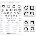
Tests that use simple line drawing pictures, instead of letters or numbers, as targets to measure a young child's visual acuity.
Preferential Looking Test
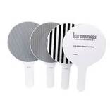
This is a visual acuity test for nonverbal children that uses black and white striped patterns. As the stripes get thinner (and thus more difficult to see), an evaluator observes to see if a child continues to look toward the stripe patterns, an indication that the child can still see them. An acuity measure is then calculated.